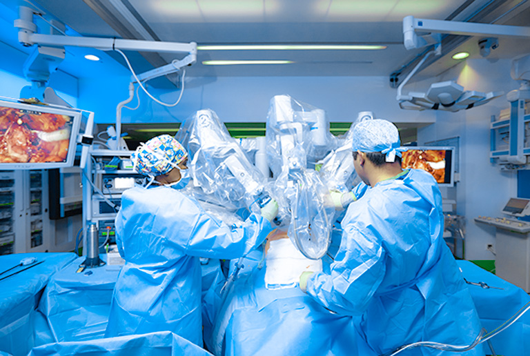
Robotic Esophagectomy
The robotic esophagectomy is a surgical operation used to remove the cancerous portion of the oesophagus, which is the foot-long conduit that connects the back of the throat to the stomach. Robotic surgeon in hyderabad is being utilised more often to treat this condition because it allows the intra-abdominal oesophagus and stomach to be completely mobilised without the need for a large abdominal incision, which leads to reduced post-operative discomfort and scarring and a speedier recovery. Laparoscopic and thoracoscopic esophagectomy techniques have limits, which robotic technologies allow surgeons to overcome.
As a result, there may be a reduction in intraoperative problems during mediastinum oesophageal dissection. It enables three-dimensional imaging, improved magnification, and a broader range of motion of the instrument. For esophagectomy, robotic technology is being used at the Uma cancer centre by best robotic surgeon in hyderabad. Robotic methods may be used during the thoracic dissection of the oesophagus, gastric mobilisation, and intrathoracic anastomosis. Additionally, it can be accomplished while using thoracoscopic, laparoscopic, or laparoscopic hand-assisted techniques. Three 8 mm ports are positioned in the right and left subcostal regions for the robotic abdominal approach.
Supraumbilical is where the camera is inserted, and a 10 mm assistant port is put in the right paramedian location. The laparoscopic approach employs identical placements with 5 mm ports and cameras. Using two additional 5-mm trocars, the left subcostal and left paramedian locations are accessed. Both procedures remove an additional incision by bringing the feeding jejunostomy out through a port location on the left. During the procedure, three to four tiny keyhole incisions, each measuring about half an inch long, are created in the upper abdomen, chest, or lower neck. The surgeon views the surgery site using a laparoscope while operating with small computer-assisted robotic equipment on a magnified, 3D display.
Robotic surgery by robotic surgeon in hyderabad reduces postoperative pain, wound infections, ventilator reliance, cardiopulmonary issues, ICU and hospital stays, and death rates by doing away with the necessity for thoracotomy, laparotomy, or both.

Robotic Gyane Oncology Surgery
Advanced surgeon-operated robotic surgical instruments enables the less invasive removal of cancer-bearing organs and tissues during the treatment of gynaecological malignancies. Gynecological oncologists were among the first to recognise that minimally invasive surgery had a lower surgical morbidity rate and a quicker postoperative recovery. Traditional laparotomy can now be replaced by robotic surgery. Laparoscopic surgery and robotic surgery are very different from one another. During a traditional laparoscopy, pictures captured by a two-dimensional camera are projected onto monitors inside the operating room and placed close to the robotic surgeon.
Through abdominal ports and 5- to 12-millimeter incisions, the surgeon performs surgery while being directly assisted at the operating table by a camera and stiff equipment. Limitations of conventional laparoscopy are frequently addressed.
Robotic surgery for cervical cancer
Many gynecologic oncologists are still unsure about whether robotic-assisted minimally invasive surgery can completely replace traditional laparotomy in all gynecologic cancer patients. According to the available research, robotic surgery can effectively diagnose cervical cancer patients’ pathological tumour size, tumour grade, deep cervix organ invasion, lymphovascular invasion, cancerous lymph node status, and cancer-free resection margins without the need for unnecessary intraoperative resection.
Robotic surgery for endometrial cancer
Robotic surgery has emerged as the current gold standard for the surgical treatment of endometrial cancer. Gynecologic oncologists quickly realised the advantages of robotic-assisted surgery for endometrial cancer patients. Initial studies complimented the use of straightforward surgical procedure, the suitability of surgical specimens for cancer staging, and the shortening of patient hospital stays and recovery times. The surgical faith insufficient lymphadenectomy (i.e., > 4 lymphadenectomies) was a fundamental advantage of the robotic platform.
Robotic surgery for ovarian cancer
Treatment for epithelial ovarian cancer centres on cytoreductive surgery, which leaves fewer than 1 cm of residual disease. Since the 1990s, efforts have been made to maximise ovarian cancer cytoreduction by minimally invasive techniques. Whether robotic-assisted surgery improves the likelihood of surgically minimising ovarian cancer is an unanswered topic. The inability to concurrently operate in the pelvis and abdomen is one of the primary drawbacks of the robotic platform, despite the possibility that enhanced ergonomics may help in radical surgery of this nature. It is an alternative for ovarian cancer in its early stages. Unless future study shows otherwise, robotic surgery is generally not advised for the treatment of ovarian cancer.
Our gynaecological oncologists may use the robotic platform to perform a significant surgery in a minimally invasive manner. One huge incision is not necessary since just a few small ones are needed to make room for the robotic tools that have been shrunk down in size and the tiny camera that has been placed into the patient’s belly. These are still essential surgical operations, but because they are performed through tiny incisions, there is far less bleeding, pain, scarring, and risk of infection than with open surgery.
